Glaucoma Awareness: What You Need to Know
January is Glaucoma Awareness Month: Did you know that 2.7 million people in the United States alone have Glaucoma? Of those 2.7 million, only 50% know they even have it! Glaucoma is often called the “Silent Thief of Sight” because early symptoms don’t present clearly in terms of vision loss. So what do you need to know about Glaucoma? Read on to learn more!
What is Glaucoma?
Glaucoma is a group of eye diseases that lead to the progressive damage of the optic nerve. The optic nerve is a bundle of nerve fibers that connect the retina, the eye structure responsible for detection of images, to the brain. The most common form of glaucoma, open angle glaucoma, is thought to be caused by an increase in pressure inside the eye that eventually leads to damage of the nerve fibers in the optic nerve. This is why your eye care professional always performs that pressure test with the puff of air at your yearly eye checkups! There is no cure for glaucoma, and vision loss from the disease cannot be restored.

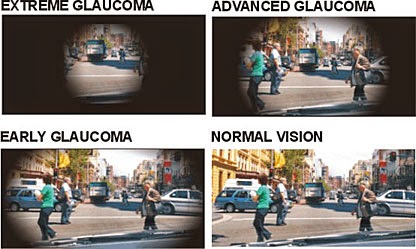
In a healthy eye, the clear fluid that fills the anterior chamber, which is the space between the cornea and the iris, flows in and out continuously helping to nourish the nearby tissues. This fluid leaves the chamber through a spongy meshwork, like a drain, that is located in the open angle of the chamber, where the iris and cornea meet. In open angle glaucoma, the fluid passes too slowly through the drain, even though the angle is wide open, and this causes the fluid to build up in the eye, increasing the pressure. This increase in pressure can damage the nerve fibers which results in vision loss. Glaucoma vision loss is initially detected in the periphery. Patients lose their side vision first, and, if it progresses further without treatment, patients then develop tunnel vision resulting in blindness. Many studies have shown that controlling the pressure inside the eye can help reduce the risk of optic nerve damage.
Whether you develop glaucoma or not depends on the amount of pressure your optic nerve can withstand without being damaged. Everyone’s eyes are different–a high pressure for one patient may be a normal pressure for another. It is important to see your eye doctor each year for a comprehensive dilated eye exam so that they can determine what a normal eye pressure is for you.
How is Glaucoma Detected?
Glaucoma is detected during a comprehensive dilated eye exam. Changes in the appearance of the optic nerve over time, loss of nerve tissue, and corresponding loss of vision are some of the factors that inform a diagnosis of glaucoma. Early detection is key to success when treating glaucoma.
A comprehensive eye exam to determine if a patient has glaucoma will include:
- Visual Acuity: to record your best corrected vision and discover if vision is affected
- Visual Field: to measure any loss of peripheral vision
- Tonometry: to measure the pressure inside the eye
- Pachymetry: to measure the thickness of your cornea. A thinner cornea may increase the risk of developing glaucoma.
- Evaluation of the Retina: a dilation drop is placed in each eye to make your pupils larger so the eye doctor can get a full view of your retina and optic nerve to assess for damage.
- Gonioscopy: to view the angle structures where the fluid drains in the eye
How is Glaucoma Treated?
Early treatment is essential for a good prognosis of the disease. The goal of treatment is to save any remaining vision, as sight that is lost from optic nerve damage cannot be reversed. The most common treatment for glaucoma is a medication in the from of an eye drop. These eye drops work to keep the pressure low in order to limit the amount of damage done to the nerves. These drops must be used regularly and consistently for optimal effect. Because glaucoma does not have clear symptoms, it is easy to forget to use the medications or simply decide to stop taking them. If you stop using the drops without the consent of your eye doctor, then the pressure in your eyes will rebound and increase to levels that will damage your optic nerve once again.
Another treatment option is Laser Trabeculoplasty. This procedure is performed after numbing drops are instilled in each eye. The doctor then uses a laser and special lens to create holes in the drainage meshwork of the angle to allow the fluid to drain easier.
If these two treatment options do not alleviate the pressure in the eye, other courses of action may have to be taken. Conventional surgery is performed to create a new drainage area so the fluid can accumulate and drain into the vascular system. This called a Trabeculectomy. Drainage implants, which are small silicone tubes, can be inserted into the eye to help drain the fluid from the anterior chamber as well. These implants are typically used for people with uncontrolled glaucoma or in children who have glaucoma.
Acute Angle Closure Glaucoma
Acute angle closure glaucoma is a medical emergency. This occurs when the fluid in the anterior chamber cannot drain through the angle because it is blocked by a part of the iris. This causes an immediate increase in pressure, leading to severe pain, nausea, redness, and blurred vision. If this occurs, seek immediate medical attention.
Life-Long Treatment
Patients with glaucoma will need to continue treatment for the rest of their lives, as there is no cure for the disease. Compliance with medications and regular eye examinations are essential, as treatment may need to be adjusted from time to time. By keeping the eye pressure under control, damage to the optic nerve may be halted and vision loss will cease to progress.
Whether you are at risk for glaucoma due to family history or not, regular eye examinations are key to detecting this disease that too often catches patients by surprise, and many other serious eye conditions that present no obvious symptoms.